
Urgent Overview: What Owners Should Do Right Now
If you suspect your dog has a gastrointestinal (GI) obstruction, this is an emergency. Intestinal blockages, which may involve a complete or partial blockage of the intestinal lumen, can become life-threatening within 24–72 hours, and waiting to see if symptoms improve on their own can be fatal.
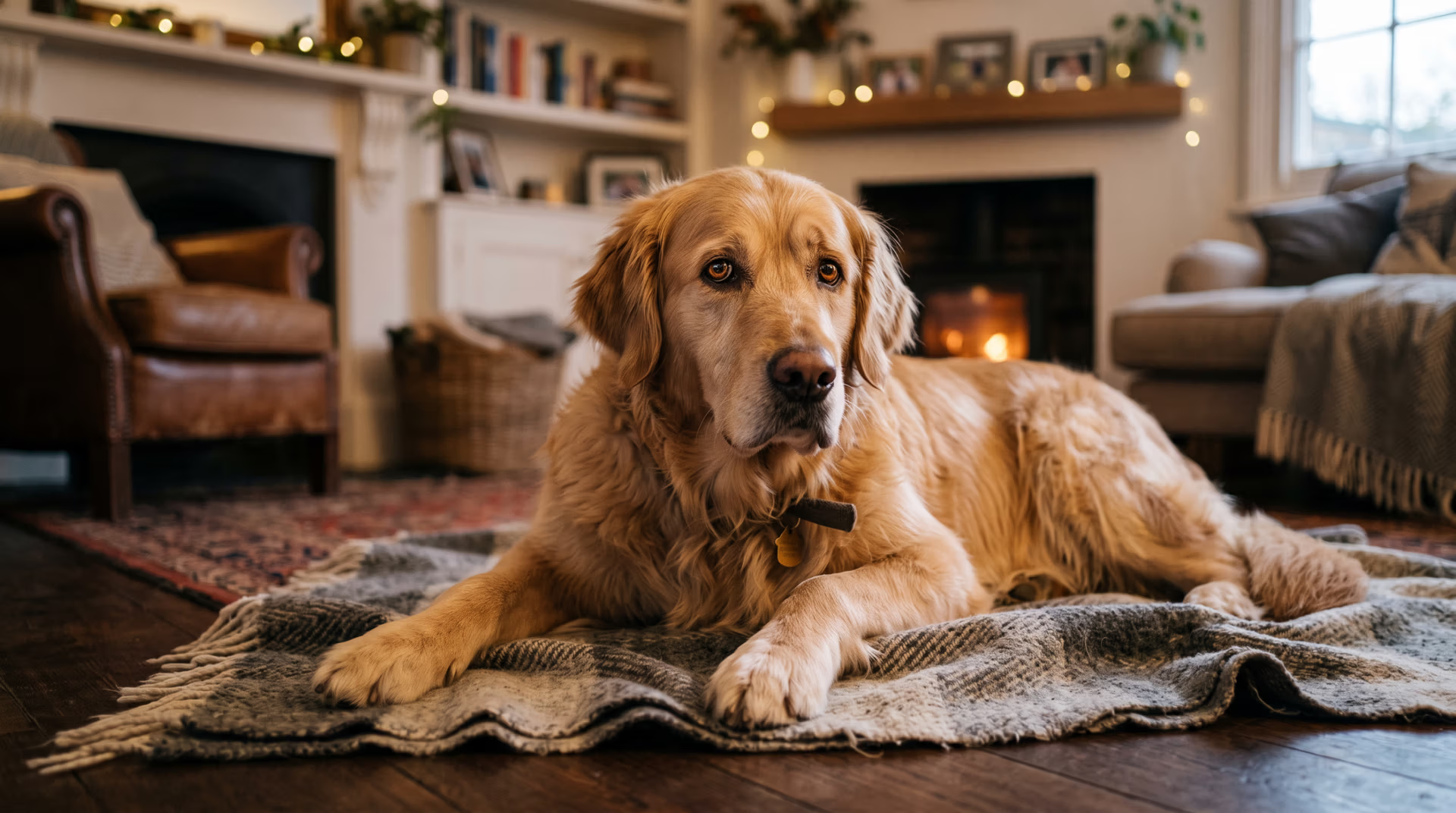
The red-flag signs you need to recognize: repeated vomiting (with or without food), sudden lethargy, a painful abdomen, and changes in bowel movements—whether that means no stool at all or only small amounts of diarrhea. Dogs with a complete blockage often cannot keep even water down. Severe symptoms of gastrointestinal obstruction include repeated vomiting, loss of appetite, lethargy, abdominal pain, and inability to defecate.
If you saw your dog swallow a foreign object—a sock, corn cob, bone, rock, string, toy, or peach pit—or if your dog has persistent vomiting and cannot hold down water, call an emergency veterinarian or 24/7 clinic immediately. Do not take a “wait and see” approach.
Here’s what you should avoid doing before speaking to a vet:
Do not induce vomiting at home without veterinary guidance
Do not give human medications (ibuprofen, Pepto-Bismol, etc.)
Do not feed food, oil, or bread hoping to “push” the object through
Do not attempt to pull string or fabric from your dog’s throat or rectum
Some objects—batteries, sharp bones, caustic materials, or linear foreign bodies like string—can cause far worse damage if vomiting is induced or if they’re pulled. Your veterinarian or poison control can advise you on the safest next step.
Early treatment often means shorter surgery, lower overall cost, fewer complications, and a dramatically better survival rate compared with dogs that arrive after 2–3 days of symptoms. In the sections that follow, I will walk both dog owners and primary care vets through causes, diagnostics, treatment options (including intestinal surgery), and realistic outcomes based on my experience as a veterinary surgeon.

What Is a Gastrointestinal (GI) Obstruction in Dogs?
A gastrointestinal obstruction—also known as an intestinal obstruction, gastrointestinal blockage, or intestinal blockage in dogs, or bowel obstruction in dogs—is a partial or complete blockage anywhere along the digestive tract, most commonly in the stomach or small intestine, that prevents the normal passage of food, fluid, and gas. When material cannot move through, it accumulates upstream, causing distension, pain, and progressive damage to the intestinal wall.
You may hear this condition called:
GI obstruction
Bowel obstruction
Intestinal blockage
Foreign body obstruction
These terms are often used interchangeably, though the most dangerous scenarios typically involve the stomach and small intestines rather than the colon.
Why obstruction is so dangerous
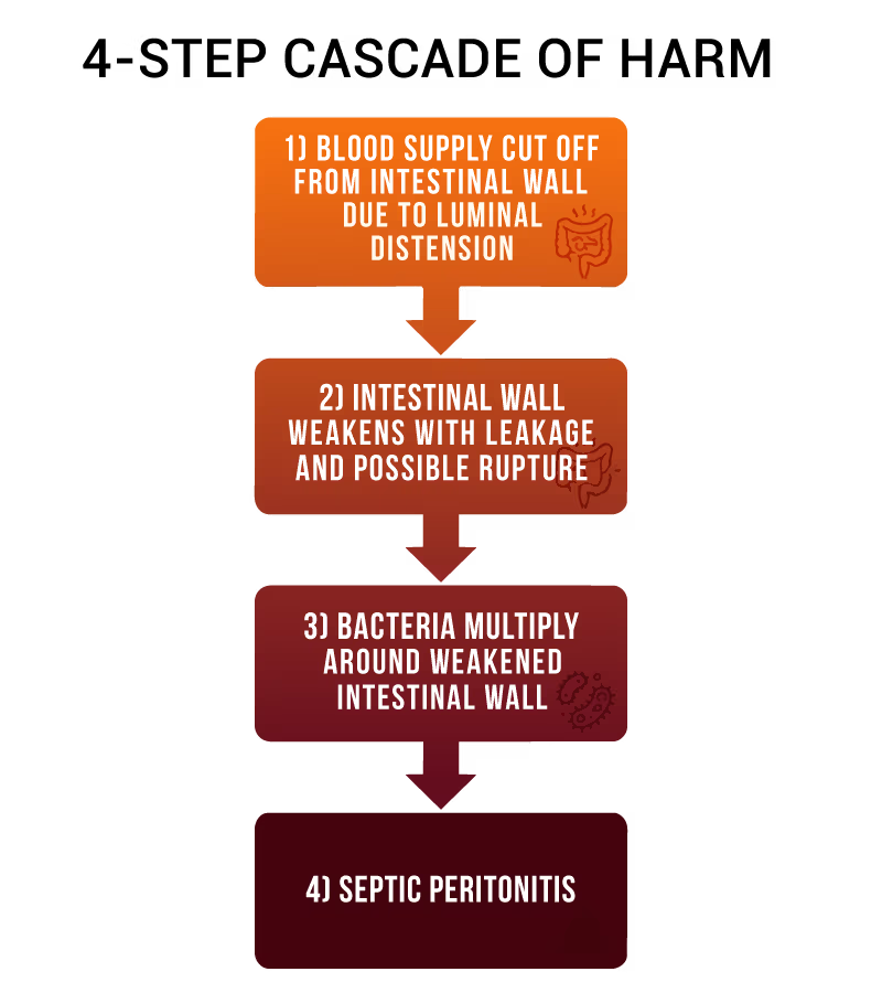
The blockage itself is only part of the problem. When the gastrointestinal tract becomes obstructed, distension and pressure can impair the intestine’s blood supply. Obstruction can also cause decreased blood flow, raising the risk of tissue necrosis, perforation, and septic peritonitis.
. Signs of gastrointestinal obstruction can vary depending on the location and severity of the blockage, which affects symptoms such as vomiting and abdominal pain.
| What Happens | Why It Matters |
|---|---|
| Blood supply to the gut wall is compromised | Decreasing blood flow leads to tissue death (necrosis) |
| The intestinal wall weakens and may rupture | Bowel rupture allows bacteria to leak into the abdominal cavity, causing septic peritonitis |
| Fluids pool in the intestines instead of being absorbed | Severe dehydration and electrolyte imbalances develop rapidly |
| Bacteria overgrow in the stagnant intestinal contents | Toxins can translocate into the bloodstream, leading to blood poisoning |
Even a partial blockage is unstable. It may progress to a complete obstruction at any time and should be monitored very closely—or surgically explored if the dog is deteriorating.
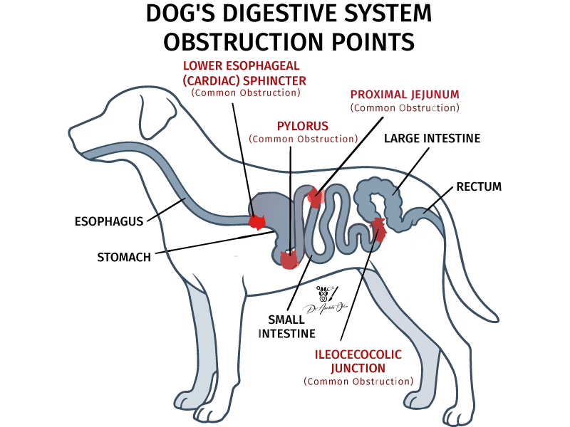
For veterinarians: High-risk anatomic locations include the pylorus, duodenal flexure, proximal jejunum, and ileocecocolic junction. Linear foreign bodies are particularly dangerous because they anchor at one point (often the stomach or base of the tongue) while peristalsis causes the intestine to plicate, resulting in plication injuries and multiple serosal perforations along the mesenteric border.
Common Causes of GI Obstruction in Dogs
While foreign body obstruction is the most common cause in young dogs, common bowel obstructions are a frequent health concern in dogs and can result from a variety of factors. Older dogs are more likely to develop obstructions from intestinal tumors or chronic inflammatory conditions. Other common causes of bowel obstruction include masses, tumors, or other issues that block the digestive tract, and other common bowel obstructions may develop when growths narrow different parts of the digestive tract. Gastric outflow obstruction is another important upper-GI form seen in dogs Intestinal obstructions in dogs can also be caused by several conditions, such as inflammation or parasites. Understanding the cause helps guide treatment and prognosis, .
Foreign Bodies: The Most Common Culprit
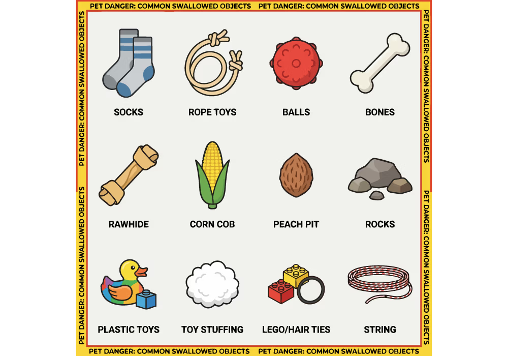
Dogs are naturally curious animals, and many will swallow just about anything. When a dog ingests foreign objects, these items can become lodged in the dog’s intestines, leading to dangerous blockages that may require emergency surgery. The objects I see most frequently during intestinal blockage surgery include:
Socks, underwear, and towels
String and rope toys (especially when shredded)
Balls and rubber toys too small for the dog’s size
Bones (raw and cooked)
Rawhide chews
Corn cobs
Peach pits, avocado pits, and similar fruit seeds
Rocks and pebbles
Plastic chew toys and fragments
Stuffing from plush toys
Children’s toys (Lego pieces, small figurines, hair ties)
Linear Foreign Bodies: A Special Danger
Linear foreign bodies—string, rope, tinsel, dental floss, yarn, and fabric strips—deserve special mention because they’re uniquely destructive. One end often anchors under the tongue or in the dog’s stomach while the rest passes into the intestines. As peristalsis tries to move the material through, the intestine bunches up like an accordion on a clothesline.
The taut string acts like a saw, cutting through the intestinal wall, resulting in multiple perforations and widespread peritonitis. These cases often require extensive surgery with multiple enterotomies or resections.
Non–Foreign Body Causes
Not every bowel obstruction in dogs involves something the dog ate. Obstructions can occur in the dog’s stomach and intestines, caused by a variety of conditions. Other causes include:
| Cause | Typical Patient |
|---|---|
| Intestinal tumors (adenocarcinoma, lymphoma, leiomyosarcoma) | Dogs over 8–9 years old |
| Pyloric stenosis or gastric outflow obstruction | Young to middle-aged dogs, especially brachycephalic breeds |
| Intussusception (telescoping of bowel) | Puppies with enteritis or parasites |
| Strictures and scar tissue | Dogs with prior abdominal surgery or severe inflammation |
| Adhesions | Dogs with previous surgery or peritonitis |
| Hernias with entrapped bowel | Intact males (perineal hernia), trauma cases |
| Mesenteric torsion or volvulus | Any age, often acute and catastrophic |
Large-breed, deep-chested dogs can develop gastric dilatation-volvulus (GDV), a related but distinct emergency where the stomach twists on itself and obstructs outflow. GDV requires immediate emergency surgery and is almost always fatal without intervention.
Age Patterns
Young dogs (especially under 3 years): Most prone to ingesting non food material like toys, socks, and rocks
Senior dogs (over 8–9 years): More likely to obstruct from intestinal tumors or strictures from chronic disease
Signs and Symptoms of GI Obstruction in Dogs
Clinical signs vary depending on where the blockage is located and how long it has been present. However, persistent vomiting is the most common early warning sign I see.
In veterinary medicine, it is important to consult a veterinarian if you notice any concerning symptoms. Veterinarians monitor dogs for visible bloating, straining to defecate, and excessive drooling as signs of gastrointestinal obstruction.
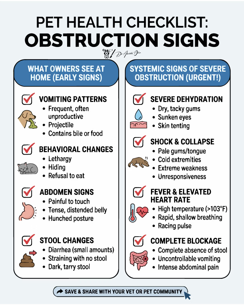
What Owners Can Recognize at Home
Vomiting Patterns
Repeated vomiting—food, foam, or yellow bile
Vomiting shortly after eating or drinking
Inability to keep water down
Drooling or hypersalivation between vomiting episodes
Behavioral Changes
Reluctance to eat or complete loss of appetite
Hunched posture or arched back
Restlessness, pacing, or inability to get comfortable
Hiding or withdrawing from family members
Refusal to lie down normally
Signs of a Painful Abdomen
Crying, yelping, or growling when the belly is touched
Rigid or tense abdominal muscles (guarding)
“Prayer position”—front legs stretched forward, back end raised
Aggression when you attempt to touch or pick up the dog
Stool Changes
No bowel movements at all
Straining to defecate with minimal or no result
Passing only small amounts of diarrhea or mucus
Tarry, dark stools (melena) suggesting bleeding
If you are wondering whether a dog can pass a foreign object (sometimes called a ‘dog pass’), it depends on the object’s size and type. Veterinary intervention is often necessary, and diagnostic tools such as X-rays or endoscopy may be required to determine whether your dog can safely pass the obstruction.
Important: Some dogs with a partial blockage will still pass stool or have diarrhea, so the presence of poop does not rule out obstruction. If your dog’s symptoms include ongoing vomiting and poop with a painful abdomen, you still need urgent veterinary evaluation.
Symptoms of a dog’s intestinal blockage generally include vomiting and listlessness.
Systemic Signs of Severe Obstruction
When obstruction has been present for more than 24 hours—or when perforation has occurred—you may see:
Profound lethargy or weakness
Rapid heart rate
Pale, gray, or muddy gum color
Collapse or inability to stand
Cold extremities
Signs of shock
These systemic signs indicate the dog is critically ill and needs emergency care immediately.
The “Upset Stomach” Trap
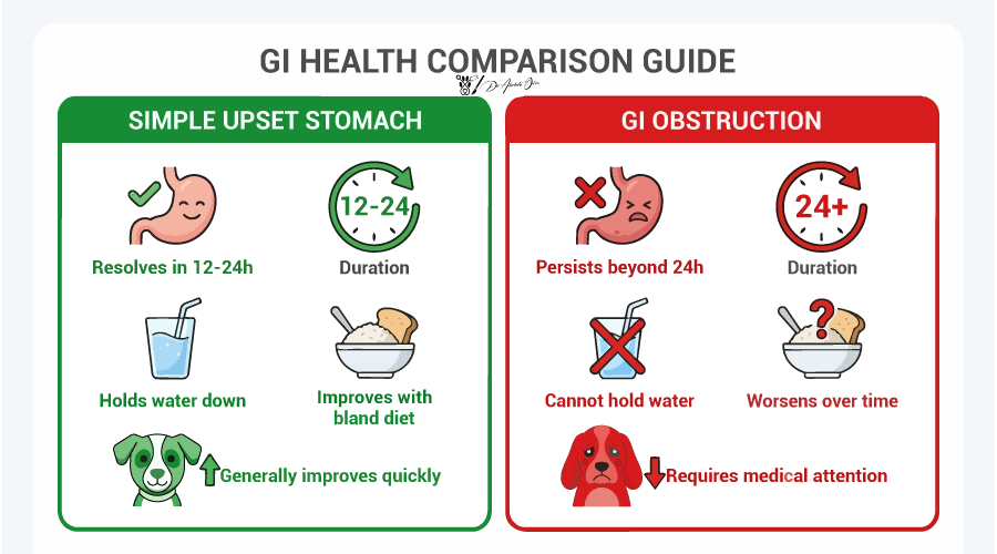
Dog intestinal blockage symptoms can initially mimic a simple upset stomach or dietary indiscretion. The critical difference is persistence. An upset stomach typically improves within 12–24 hours with rest and a bland diet. Obstruction does not improve—it gets worse.
When to worry:
Vomiting continues beyond 12–24 hours
Dog cannot keep water down
Any sudden severe abdominal pain
Known or suspected ingestion of a foreign object
How Veterinarians Diagnose GI Obstruction
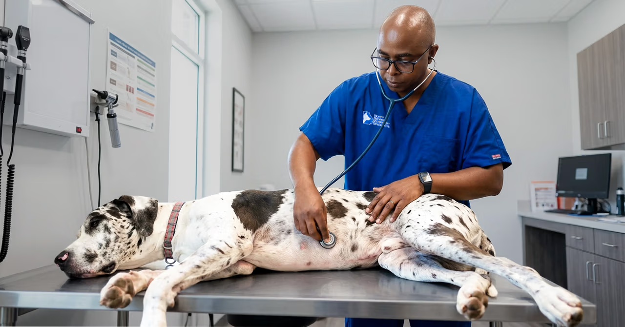
Diagnosing dog intestinal blockages involves history-taking, physical exam findings, blood work, and imaging. Many veterinary clinics have an in-house diagnostic lab where advanced imaging and tests, such as X-rays and ultrasounds, can be performed on-site to quickly diagnose issues like foreign object ingestion. Diagnosis of gastrointestinal obstruction involves a combination of veterinary history, physical examination, imaging techniques such as X-rays and ultrasound, and bloodwork. Each piece of information helps build the clinical picture.
History and Physical Exam
The first questions I ask owners:
Did you see the dog eat something unusual?
When did symptoms start?
How often is the dog vomiting?
Has the dog passed stool? What did it look like?
Any previous diet changes or garbage access?
History of eating foreign objects?
Even if you didn’t witness your dog swallowing something, a negative history doesn’t rule out foreign body obstruction. Many dogs ingest objects when owners aren’t watching.
The physical exam evaluates:
Hydration status (skin turgor, gum moisture, capillary refill time)
Heart rate and pulse quality
Abdominal palpation—feeling for gas-distended loops, painful segments, or a discrete mass
Rectal exam if indicated
Temperature (fever may suggest perforation; hypothermia suggests shock)
In some cases, an endoscopy may be performed by passing a camera through the dog’s throat to examine the stomach for foreign objects or blockages.
When Inducing Vomiting Might Be Considered
If ingestion of a foreign object occurred within the last 30–120 minutes and the dog is otherwise stable, I may consider inducing vomiting—but only after confirming it’s safe. Vomiting is contraindicated when:
The object is sharp (bones, needles, glass)
The material is caustic (batteries, certain household chemicals)
It’s a linear foreign body
The dog is already vomiting, seizing, or has diminished consciousness
Never induce vomiting at home without veterinary guidance.
Imaging: X-Rays and Ultrasound
Abdominal radiographs are typically the first imaging step. We look for:
Gas patterns—two populations of intestinal loops (some normal, some dilated)
“Stacking” or “hairpin” patterns suggesting chronic obstruction
Visible radiopaque foreign objects (metal, bones, rocks)
Plication patterns with linear foreign bodies
Loss of serosal detail or free gas indicating perforation
However, many foreign objects—fabric, plastic, rubber—don’t show up on X-rays. That’s where ultrasound becomes invaluable.
Abdominal ultrasound can:
Identify non-radiopaque foreign bodies
Assess intestinal wall thickness and layering
Detect intussusception (classic target or bull’s-eye sign)
Evaluate for free abdominal fluid
Guide abdominocentesis if peritonitis is suspected
For an accurate diagnosis, I often recommend both modalities.
Blood Work
A complete blood count (CBC) and serum chemistry panel help evaluate:
Dehydration and kidney function (BUN, creatinine)
Electrolyte imbalances (hypochloremia and hypokalemia with proximal obstruction)
White blood cell changes suggesting infection or sepsis
Lactate levels indicating tissue hypoperfusion
Low albumin, which increases surgical risk
This information guides fluid therapy, anesthesia planning, and prognosis.
For Veterinarians: When Imaging Is Equivocal
In uncertain cases, options include:
Serial imaging after 4–6 hours to assess progression
Contrast studies (barium or iodinated contrast)
CT scan if available
Early exploratory laparotomy when clinical suspicion is high, and the patient is deteriorating
When in doubt—especially in a vomiting dog that isn’t improving—I favor surgical exploration over continued monitoring. The intestine doesn’t get healthier with time.
Treatment Options for GI Obstruction in Dogs
Complete obstructions almost always require urgent intervention—endoscopy or surgery. Treatment for gastrointestinal obstruction often involves IV fluids, surgical removal of the foreign object, and supportive care. “Watchful waiting” is reserved only for very carefully selected, stable patients with partial obstructions under close veterinary supervision.
Conservative Management
In specific situations, medical management may be attempted:
The object is small, smooth, and already in the large intestine on imaging
The dog is clinically stable with no signs of obstruction or pain
Serial imaging shows the object is moving through the GI tract
Conservative dogs treatment includes:
IV fluids to prevent dehydration
Anti nausea medications (maropitant/Cerenia)
Pain medications as needed
Serial abdominal radiographs every 4–12 hours
If the object fails to progress, or if the dog’s symptoms worsen, surgery becomes necessary. Conservative management requires hospitalization and close monitoring—it’s not appropriate for at-home “wait and see.”
Endoscopic Retrieval
When the foreign object is in the esophagus, stomach, or proximal duodenum, endoscopy may be an option. The dog is anesthetized, a flexible camera is passed down the dog’s throat, and grasping tools are used to retrieve the object without an abdominal incision.
Advantages of endoscopy:
Minimally invasive
Shorter recovery time
No abdominal incision
Limitations:
Cannot reach objects in mid or distal small intestine
Not appropriate for linear foreign bodies (pulling can cause more damage)
Sharp objects may injure the esophagus during retrieval
Requires specialized equipment and expertise
When Surgery Is Necessary
Surgical removal is recommended when:
The object is located in the small intestine beyond endoscopic reach
Linear foreign body is confirmed or suspected
There is evidence of perforation or peritonitis
Endoscopic retrieval fails or is contraindicated
The dog is deteriorating despite medical management
Complete intestinal blockage is confirmed
In these cases, bowel obstruction surgery is necessary to remove the obstruction. Intestinal blockage surgery for dogs is a major procedure that requires anesthesia, and whether a dog recovers well from it—intestinal blockage surgery—depends on how early the problem is diagnosed, how much bowel is damaged, and the dog’s overall condition.
The timing equation is simple: Delaying surgery in unstable or clearly obstructed patients dramatically increases the risk of intestinal rupture, septic peritonitis, and death. I’ve seen dogs that were stable one morning become critical by that afternoon. The intestine doesn’t wait.
Surgical Management of GI Obstruction and Gastrointestinal Blockage Surgery Cost
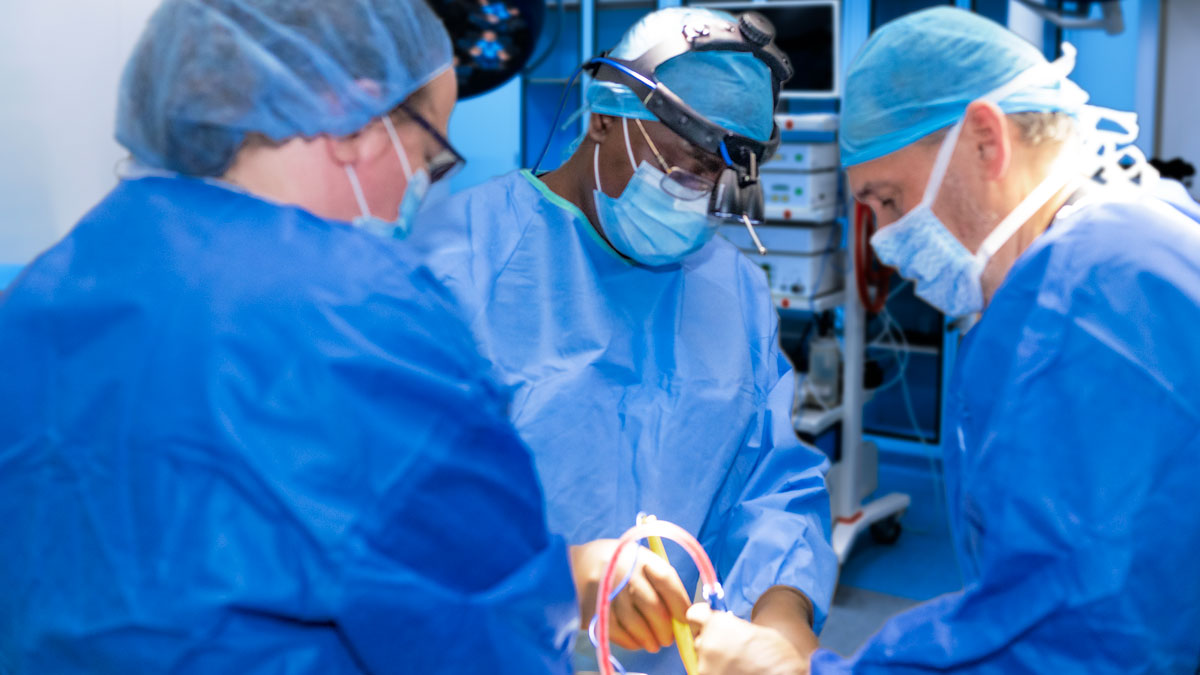
Intestinal blockage surgery is performed under general anesthesia through a midline abdominal incision that allows access to the entire GI tract from the stomach to the colon. A dog’s intestinal blockage surgery is a critical procedure that not only removes the obstruction but also helps prevent life-threatening complications. The cost of intestinal blockage surgery varies with procedure complexity, the dog’s health status, and geographic location, so owners should ask their veterinarian for an estimate tailored to their dog’s needs.
Pre-Surgical Stabilization
Before anesthesia, most dogs need:
IV fluid resuscitation to correct dehydration
Electrolyte correction
Anti-nausea medications
Pain control (typically opioid-based)
Broad-spectrum antibiotics if perforation is suspected
Stabilization time depends on severity. A mildly dehydrated dog may need only an hour of fluids; a dog in septic shock may require several hours of intensive care before becoming a reasonable candidate for anesthesia.
Common Surgical Procedures
Gastrotomy: Opening the dog’s stomach to remove an object lodged there. The stomach is isolated, incised, the foreign material is extracted, and the stomach is closed in layers. I always examine the pylorus and run the small intestine to check for additional objects.
Enterotomy: Opening the small intestine to remove a foreign body. An incision is made on the anti-mesenteric border near the obstruction, the object is removed, and the intestine is closed. This is the most common procedure for foreign body obstruction.
Resection and anastomosis: If a segment of the dog’s intestines is necrotic—dark, thin, lacking blood flow—that portion must be removed and the healthy ends sutured together. This is more complex surgery with higher post-operative risk but is necessary to prevent continued tissue death and perforation.
Intraoperative Decision-Making
During surgery, I assess each intestinal segment for:
Color (pink and healthy vs. dark purple or gray)
Wall thickness
Arterial pulsations in the mesentery
Peristalsis when touched
If viability is questionable, resection is safer than leaving compromised bowel that may perforate later.
For linear foreign bodies, multiple enterotomies along the length of intestine are often necessary. These cases frequently involve serosal tears at the mesenteric border that must be carefully sutured.
When Perforation Has Occurred
In cases with free abdominal fluid or confirmed bowel rupture, the abdominal cavity is thoroughly lavaged with warm sterile fluids. A feeding tube (esophagostomy or jejunostomy) may be placed if prolonged recovery is anticipated. A closed suction peritoneal drain may also placed to prevent accumulation of inflammatory or infected fluid within the abdominal cavity.
Hospitalization After Surgery
Typical post-operative care includes:
Continued IV fluids until the dog is drinking on their own
Post-surgery pain medication (opioids transitioning to oral)
Anti-nausea therapy
Monitoring of temperature, heart rate, abdominal comfort, and incision
Expected hospitalization:
Straightforward foreign body removal: 1–3 days
Perforation or extensive resection: 3–5+ days
Recovery, Prognosis, and Potential Complications
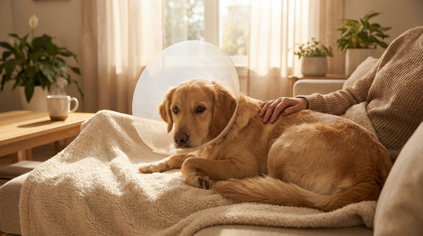
Prognosis is generally excellent—often over 90% survival—for otherwise healthy dogs treated early, before the intestine becomes necrotic or perforates. Managing your dog’s pain during recovery from intestinal blockage surgery is crucial, as appropriate pain medications and monitoring can help ensure a smoother healing process. The longer the delay, the worse the outcome.
What to Expect After Discharge
Enforce 10–14 days of strict rest (short leash walks only for bathroom breaks)
Use an Elizabethan collar to protect the incision at all times
Feed small, frequent meals of a bland or prescription GI diet
Administer medications as directed (pain medications, anti-nausea, sometimes antibiotics)
Keep the incision clean and dry
Warning Signs That Require Urgent Recheck
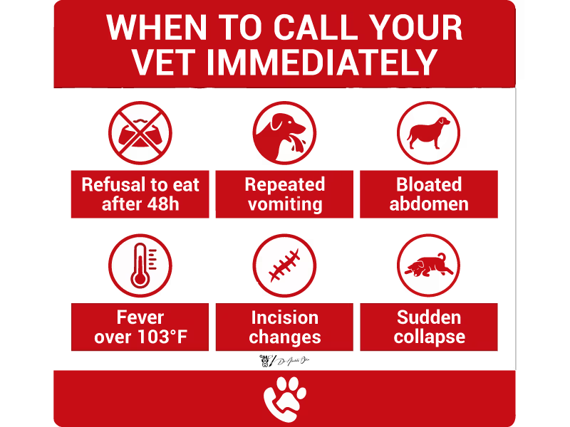
Contact your veterinarian immediately if you observe:
Refusal to eat after 24–48 hours at home
Repeated vomiting
Bloated, distended, or painful abdomen
Fever (rectal temperature over 103°F)
Incision swelling, redness, discharge, or gaping
Sudden lethargy, weakness, or collapse
Potential Complications
The critical window for complications is days 3–5 after surgery, when intestinal healing is weakest relative to mechanical stress. Complications include:
| Complication | What It Means |
|---|---|
| Surgical site infection | Incision becomes red, swollen, or drains pus |
| Dehiscence | Intestinal sutures fail, causing leakage and septic peritonitis—this is a surgical emergency |
| Short-bowel syndrome | After major resection, diarrhea and malabsorption may occur |
| Adhesions | Scar tissue forms between intestinal loops, potentially causing future obstructions |
Recovery Timelines
Uncomplicated cases: Return to normal activity by 2 weeks
Cases with perforation or extensive resection: Several weeks of gradual improvement with close veterinary follow-up
Dogs recovering from septic peritonitis: May require weeks to months of recovery; some do not survive despite aggressive treatment
Timing matters: The dog intestinal blockage timeline before treatment is measured in hours to days, while recovery after successful surgery ranges from about 2 weeks in uncomplicated cases to much longer when perforation or sepsis occurred
Preventing Intestinal Blockages and How to Reduce the Risk of GI Obstruction
Prevention is far easier, safer, and less expensive than emergency surgery—especially for adventurous puppies and heavy chewers. As a surgeon, I’d much rather help you prevent intestinal blockages than remove them.
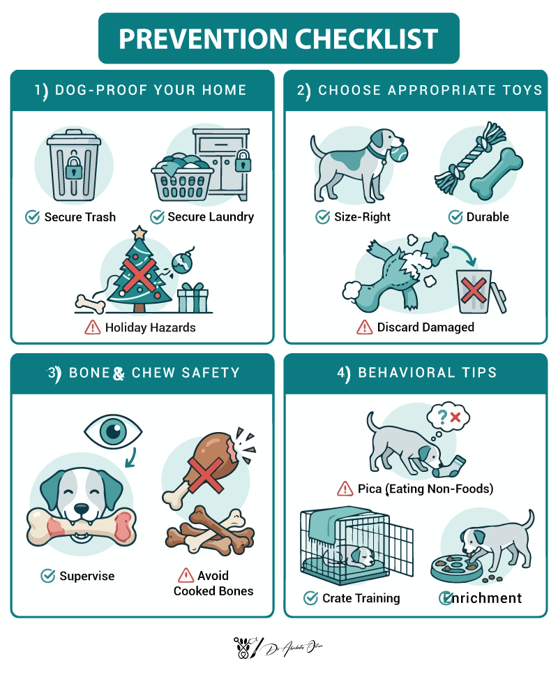
Dog-Proof Your Home
Keep laundry (especially socks and underwear) in closed hampers
Use trash cans with secure lids
Store children’s toys, hair ties, rubber bands, and craft supplies out of reach
Be mindful of holiday hazards: tinsel, ribbon, ornament hooks
Secure any string, yarn, or rope that a dog-eat scenario could occur
Choose Appropriate Toys
Select size-appropriate, durable toys designed for your dog’s chewing strength
Avoid toys that can be shredded into long strips or small swallowable pieces
Discard damaged toys immediately—especially once stuffing becomes accessible
Supervise play with any toy that could be destroyed
Be Cautious with Bones and Chews
Supervise dogs with bones, rawhides, antlers, and bully sticks
Avoid cooked bones, which splinter easily
Remove chews once they become small enough to swallow whole
Consider alternatives like frozen Kongs or puzzle feeders for curious dogs
Supervise and Confine
Crate training provides safe confinement when you can’t supervise
Young dogs under 2–3 years need extra vigilance
“Repeat offenders” who have eaten foreign objects before are at high risk of doing it again
Address Underlying Behavior
Some dogs develop pica—the compulsive eating of non-food items—due to anxiety, boredom, or medical conditions. If your dog repeatedly ingests foreign objects despite environmental management, consider:
Consultation with a veterinary behaviorist
Increased exercise and mental enrichment
Medical workup for nutritional deficiencies or GI disease
Anxiety management strategies
Frequently Asked Questions About GI Obstruction in Dogs
Will my dog still poop if they have a blockage?
Yes, some dogs with partial blockage will pass small or thin stools, or even have diarrhea, because liquid can squeeze around the obstruction. The presence of poop does not rule out intestinal blockage. If your dog has persistent vomiting combined with any changes in stool—or a lack of stool—seek veterinary care immediately.
How long can a dog live with a GI obstruction?
This is the dog intestinal blockage timeline that concerns me most: complete blockage can become fatal within 2–4 days. Many dogs are critically ill by 24–48 hours, especially if the bowel perforates. Partial obstructions are unpredictable and can progress to complete obstruction at any time. There is no safe window for waiting at home.
Can walking or home remedies clear a blockage?
Walking may help mild constipation, but it will not fix a true mechanical obstruction. Home remedies—feeding bread, giving oil, using laxatives—are ineffective for obstruction and delay proper treatment. In some cases, they can worsen outcomes by causing additional vomiting or pushing objects into worse positions. If you suspect obstruction, see a veterinarian immediately rather than attempting home treatment.
How much does obstruction surgery cost?
Intestinal blockage surgery cost varies significantly based on location, whether it’s an emergency, length of hospitalization, and whether bowel resection is needed. In many parts of North America, expect a range of approximately $2,500–$6,000 or more. Straightforward gastrotomy or enterotomy at a general practice will be on the lower end; emergency surgery with bowel resection and ICU care at a specialty hospital will be higher. Always ask your local veterinarian or emergency clinic for an estimate based on your specific situation.
What factors affect how much an intestinal blockage depends on for treatment?
The overall treatment approach and cost for an intestinal blockage depend on: the location of the obstruction, the duration of symptoms before treatment, whether the intestine is viable or requires resection, the presence of perforation or peritonitis, and the dog’s overall health status. Early intervention almost always results in simpler surgery and better outcomes.
Should I try to remove string from my dog’s mouth or rear end?
Never pull string, ribbon, or fabric that appears to be coming from either end. If the material is anchored internally, pulling can cause the intestine to tear, leading to perforation and peritonitis. If you see material protruding, leave it in place and get to a veterinarian immediately.
Important: The information in this article is educational and does not constitute medical advice for your individual pet. Any dog with suspected GI obstruction must be evaluated in person by a veterinarian or emergency clinic as soon as possible. If you’re reading this because you’re worried about your dog right now, please call your veterinarian immediately—don’t wait.
Key Takeaways
GI obstruction in dogs is a true emergency that can become fatal within 24–72 hours
Persistent vomiting, abdominal pain, and changes in stool are the hallmark warning signs
Young dogs most commonly obstruct from foreign bodies; older dogs may obstruct from tumors
Diagnosis requires physical exam, imaging (X-rays and ultrasound), and blood work
Most complete obstructions require surgery; endoscopy is an option only for proximal foreign bodies
Early treatment dramatically improves survival rates and reduces complications
Prevention through dog-proofing, appropriate toys, and supervision is far better than treatment
If your dog shows any combination of these warning signs, trust your instincts and seek veterinary care immediately. As a surgeon, I can tell you that the dogs I worry least about are the ones whose owners brought them in early—not the ones who waited to see if things would improve on their own. Early intervention saves lives, reduces suffering, and often saves money too.