
Introduction
This comprehensive guide covers the causes, diagnosis, treatment options, prognosis, and prevention of perineal hernia in dogs. It is intended for dog owners concerned about this condition and who want to understand how it affects their pet’s health and quality of life. Perineal hernia in dogs is a potentially life-threatening condition that can significantly impact a dog’s comfort, ability to defecate or urinate, and overall well-being. Early recognition and appropriate management are crucial for the best possible outcome.
Key Takeaways
A perineal hernia is a weakness or tear in the muscles beside the anus (the pelvic diaphragm), allowing fat or organs to bulge into the perineal region, and it mainly affects older intact male dogs.
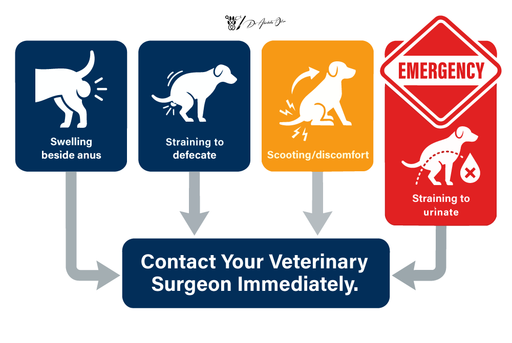
Common clinical signs include a soft swelling next to the anus, straining to pass stool or urine, constipation, and sometimes fecal incontinence.
Emergency situations occur when the urinary bladder becomes trapped in the hernia, causing sudden inability to urinate, collapse, or severe pain—this requires same-day veterinary care.
Diagnosis typically involves a digital rectal examination and imaging (radiographs or ultrasound) to identify which organs are herniated and assess urgency.
Surgical repair combined with castration in most males is the definitive treatment; outcomes are generally good with experienced surgeons, though recurrence can occur, and careful postoperative management is essential.
What Is a Perineal Hernia in Dogs?
Perineal hernia in dogs develops when the muscles that normally support the rectum and pelvic organs weaken, separate, or tear. Specifically, a perineal hernia results from weakening or complete failure of the muscular diaphragm of the pelvis. These muscles—collectively called the pelvic diaphragm—form a muscular “floor” that keeps everything in its proper place beneath the tail. When this support fails, fat, fluid, or even organs can bulge outward into the soft tissue beside the anus, creating a visible swelling under the skin.
The pelvic diaphragm is made up of several key structures: the levator ani muscle, the coccygeus muscles, and the external anal sphincter muscle. The most common perineal hernia occurs between the levator ani, internal obturator, and external anal sphincter and is termed a caudal perineal hernia. In most affected dogs, the defect occurs between these muscles, typically on one or both sides of the rectum. Surgeons often refer to this as a “caudal perineal hernia” because it sits just behind (caudal to) the main pelvic opening.
It’s important to understand that this condition is quite different from inguinal or umbilical hernias. Perineal hernias occur specifically in the region under the tail, to either side of the anus. You might notice the swelling when your dog lifts their tail or squats to defecate.
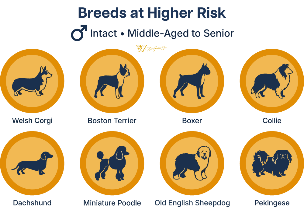
Perineal hernias occur most commonly in intact male dogs aged 5 to 7 years. However, they can also develop in neutered males, females, and occasionally in cats. Certain breeds appear over-represented in clinical reports, including Boxers, Welsh Corgis, Pekingese, and Dachshunds, though any breed can be affected.
These hernias can be classified as unilateral (affecting one side) or bilateral disease (affecting both sides). Apparently, unilateral perineal hernias occur more frequently, with the right side affected more often than the left. However, at least 30% of cases involve both sides simultaneously.
Causes and Risk Factors
Canine perineal hernia doesn’t have a single cause. Instead, it develops from a combination of hormonal influences, chronic straining, and progressive structural weakness of the pelvic muscles. Understanding these factors helps explain why certain dogs are at higher risk.
Hormonal Factors
The striking predominance of intact male dogs among affected patients strongly suggests hormonal involvement. Androgens and other hormones associated with the intact male reproductive system appear to contribute to the gradual weakening of the pelvic diaphragm muscles and connective tissue.
Concurrent prostatic disease is remarkably common—studies show that 25–69% of dogs with perineal hernia also have prostatic enlargement. An enlarged prostate causes dogs to strain when urinating and defecating, and over time, this repeated pressure stretches and fatigues the perineal muscles.
Chronic Straining
Any condition causing persistent tenesmus (straining) can contribute to the development of a perineal hernia. Common culprits include:
Chronic constipation from diet, dehydration, or megacolon
Prostatic hypertrophy causing urinary difficulty
Anal sac disease or perianal tumors
Colitis or other inflammatory bowel conditions
When dogs strain repeatedly, the muscles of the pelvic diaphragm gradually stretch, weaken, and eventually fail—much like a worn elastic band that finally gives way.
Age-Related Muscle Degeneration
As dogs age, their muscles naturally lose mass and strength. The pelvic diaphragm muscles are no exception. Combined with connective tissue changes and possibly breed-related differences in conformation, this age-related atrophy increases vulnerability to herniation.
Recognized Risk Factors
The key risk factors for developing a perineal hernia include:
Intact male status (the single strongest risk factor)
Age greater than 5–7 years
History of chronic constipation or straining
Prostatic disease (benign hyperplasia, prostatitis, cysts)
Delayed presentation after the swelling is first noticed
Breed predisposition (Boxers, Corgis, Pekingese, Dachshunds)
Importantly, comprehensive research has not supported the notion of generalized neurological or orthopedic disease as a direct cause. Any nerve changes found in the perineal region are usually secondary to the hernia itself rather than a primary cause.
Clinical Signs and When It’s an Emergency
Clinical findings in dogs with perineal hernia refer to the observed signs and symptoms that are crucial for diagnosis. These can range from subtle early changes to life-threatening emergencies. Recognizing what to watch for helps ensure your dog receives timely care.
Early Signs
The first thing most owners notice is a soft, often painless bulge beside or below the anus. This perineal swelling may:
Appear on one side or both sides
Fluctuate in size (often larger after straining)
Be reducible when gentle pressure is applied
Cause the dog no obvious distress initially
At this early stage, many dogs remain bright and active, which can lead owners to delay seeking veterinary attention.
Common Gastrointestinal Signs
As the hernia enlarges and the rectal wall becomes displaced or pouched, defecation becomes increasingly difficult:
Straining to defecate (tenesmus) with prolonged squatting
Constipation or passing only small amounts of stool frequently
Ribbon-shaped or flattened feces
Discomfort or vocalization when defecating
Occasional soiling or mild fecal incontinence
Visible rectal prolapse in severe cases
Urinary Signs
When the urinary bladder becomes involved, you may observe:
Straining to urinate with little urine produced
Longer time spent posturing to urinate
Frequent small urinations
Dribbling or urinary incontinence
Complete inability to pass urine (an emergency)
Urinary bladder retroflexion—where the bladder flips backward into the hernia—is one of the most dangerous complications and can occur suddenly.
What Can Be Trapped in the Hernia?
The contents of a perineal hernia can include:
Pelvic and retroperitoneal fat
Dilated, deviated rectum
Prostate gland
Small intestine
Urinary bladder
The clinical signs change depending on which abdominal organs have herniated and whether their blood supply or function is compromised.
Red-Flag Emergency Signs
Seek same-day veterinary care if your dog shows any of the following:
Sudden inability to urinate
A very firm, tense, or painful perineal swelling
Vomiting or retching
Collapse or severe weakness
Extreme lethargy or depression
Rapid abdominal enlargement or distension
Bladder herniation with urinary obstruction and strangulated intestinal obstruction are both life-threatening emergencies. These conditions can cause metabolic derangements, kidney failure, or intestinal necrosis within hours.
Some dogs may also carry their tails differently, excessively lick the perineal area, or show hindlimb weakness and other neurological deficits when local nerves are stretched or compressed by herniated contents rather than because of a primary neurologic cause.
How Perineal Hernia Is Diagnosed
Diagnosis of a perineal hernia is often straightforward for an experienced veterinarian, but a thorough assessment is essential to plan safe treatment and identify any emergencies that require immediate intervention.
Physical Examination and Rectal Palpation
The cornerstone of diagnosis is a careful physical examination followed by rectal examination. During rectal palpation, the veterinarian inserts a gloved, lubricated finger into the rectum to assess:
Loss of normal muscular support on one or both sides
Deviation or pouching of the rectal wall
Whether the hernia is unilateral or bilateral
What structures may be present within the defect
Prostate size and position
Many dogs require sedation or short-acting analgesia for a comfortable and thorough digital rectal examination, particularly if they are painful, anxious, or have a large hernia. The clinician may also assess for neurological deficits when nerve involvement is suspected, as these signs can reflect pressure or stretching from the hernia rather than a primary cause.
Baseline Laboratory Tests
Before anesthesia and surgery, standard laboratory evaluation includes:
| Test | Purpose |
|---|---|
| Complete blood count | Assess for infection, anemia, and hydration status |
| Serum biochemistry | Evaluate kidney function, electrolytes, and liver parameters |
| Urinalysis | Check for infection, kidney function, and urine concentration |
| Additional tests | Cardiac evaluation if indicated by age or breed |
These tests help determine anesthetic safety and identify concurrent diseases common in older dogs.
Imaging Studies
Imaging provides crucial information about hernia contents and associated conditions:
Abdominal radiographs reveal:
Position of the urinary bladder (normal vs retroflexed)
Presence of bowel loops in the hernia
Degree of fecal retention or megacolon
Prostatic size
Ultrasound helps:
Identify which specific organs are within the hernia
Measure prostate dimensions and assess for cysts or masses
Evaluate the bladder wall and other abdominal organs
CT scans may be used in referral centers for:
Complex or recurrent cases
Detailed three-dimensional mapping of the pelvic canal
Precise surgical planning
Classifying the Hernia
The diagnostic process also involves characterizing the hernia to guide surgical findings and treatment decisions:
Location (caudal, dorsolateral, ventral, or sciatic perineal hernia)
Laterality (unilateral vs bilateral)
Contents (fat only vs organ involvement)
Presence of bladder herniation or intestinal strangulation (emergency)
Concurrent prostatic disease severity
This classification directly influences which surgical technique is most appropriate and whether emergency decompression is needed before definitive repair.
Treatment Options: Medical Management vs Surgery
A perineal hernia is fundamentally a structural weakness that cannot be corrected without surgery. Medical management plays an important supportive role but should not be considered definitive
Medical Management
Medical therapy aims to:
Soften stools using high-fiber diets, prescription gastrointestinal foods, stool softeners (lactulose, psyllium), or canned pumpkin
Reduce straining by treating constipation, prostatic enlargement, or anal sac disease
Maintain hydration through oral fluids or intravenous fluids when needed
Provide pain relief and anti-inflammatory medication
Medical treatment is typically used:
While stabilizing an emergency (especially if the bladder is involved)
To prepare the colon and patient for anesthesia and surgery
When surgery must be delayed due to other health problems
As palliative care, if owners decline surgery (understanding that risks increase over time)
Without surgical correction, most hernias progressively enlarge, and the risk of life-threatening complications—particularly urinary obstruction—remains constant.
Surgical Treatment
Perineal herniorrhaphy (surgical repair of the hernia) is the standard of care. The surgical procedure involves:
Reducing herniated contents back into the pelvic canal or abdomen
Reconstructing the pelvic diaphragm using available tissues
Reinforcing the repair to prevent recurrence
Surgical Techniques
Several surgical techniques exist, with the choice depending on hernia size, tissue quality, and surgeon experience:
Traditional suture repair involves direct apposition of the remaining pelvic diaphragm muscles. This technique has higher recurrence rates and is less commonly used as a standalone approach.
Internal obturator muscle transposition is the most widely recommended technique in veterinary surgery today. The internal obturator muscle is elevated from its attachment on the pelvis and rotated to fill the hernial defect, then sutured to the external anal sphincter, coccygeus, and sacrotuberous ligament. This provides a robust, well-vascularised repair.
Other muscle flaps, such as superficial gluteal muscle transposition, may be used in complex, recurrent, or bilateral cases where the internal obturator is inadequate.
Synthetic mesh (surgical mesh, porcine dermal collagen grafts) can bridge large defects but carries a higher infection risk due to proximity to the anus and is reserved for selected cases.
Ancillary Procedures
Additional procedures are often performed during the same anesthetic:
| Procedure | Purpose |
|---|---|
| Castration | Reduces prostatic size, removes hormonal contribution, lowers recurrence risk |
| Colopexy | Fixes the colon to the abdominal wall, preventing caudal displacement |
| Cystopexy | Secures the bladder to prevent future retroflexion |
| Vas deferensopexy | Uses spermatic cord structures to anchor the bladder/prostate |
Castration at the time of perineal hernia surgery is strongly recommended in most intact male dogs. It addresses concurrent prostatic disease, reduces straining, and significantly decreases the risk of hernia recurrence and development of a hernia on the opposite side.
What to Expect From Surgery and Hospitalization
Understanding the surgical journey helps owners prepare for their dog’s treatment and recovery.
Timeline Overview
Preoperative period: Stabilization, fasting, diagnostic completion
Day of surgery: Anesthesia, perineal hernia repair, castration, any organopexies
Postoperative monitoring: Pain control, bladder management, soft diet
Hospital stay: Typically 1–3 days for uncomplicated cases
Preoperative Preparation
Before surgery, your dog will undergo:
Fasting (typically 8–12 hours) to reduce anesthetic risk
Enemas or gentle colon evacuation if significant fecal retention exists
Intravenous catheter placement for fluid therapy and medication administration
Preoperative pain relief (analgesics)
Prophylactic antibiotics to reduce surgical site infection risk
Urinary catheterization if bladder involvement is present
Anaesthesia
Modern balanced anesthesia is tailored to each patient, with continuous monitoring of:
Heart rate and rhythm (ECG)
Blood pressure
Oxygen saturation
Carbon dioxide levels
Body temperature
While anesthetic risk is higher in geriatric patients, careful planning and monitoring by experienced veterinary teams make surgery safe for most older dogs with concurrent disease.
The Surgical Procedure
In lay terms, the surgery proceeds as follows:
Your dog is positioned with the hindquarters elevated
The perineal region is clipped and surgically prepared
An incision is made beside the anus over the swelling
Herniated contents are gently returned to their normal positions
The pelvic diaphragm is reconstructed using internal obturator transposition or other techniques
Careful suture material placement avoids the sciatic nerve
Any organopexies are performed through a separate abdominal incision
Castration is completed through a prescrotal or perineal approach
All incisions are closed, and the surgical site is protected
| Technique Name | Description | Best Used When | Recurrence Risk | Notes |
|---|---|---|---|---|
| Internal Obturator Transposition | Obturator muscle elevated and rotated to close the hernia defect | Most cases; gold-standard first repair | Lowest with concurrent castration | Most commonly performed technique; preferred by soft tissue specialists |
| Superficial Gluteal Transposition | Gluteal muscle flap used to reinforce repair | Large defects; added reinforcement needed | Low when combined with primary technique | Often used in combination with internal obturator technique |
| Semitendinosus Transposition | Semitendinosus muscle flap rotated to fill defect | Recurrent hernias; failed primary repair | Moderate; depends on defect size | Alternative flap; useful when other muscles unavailable or compromised |
| Synthetic Mesh | Prosthetic mesh used to bridge or reinforce the defect | Large defects; limited autologous tissue | Variable; infection risk consideration | Select cases only; requires careful patient selection |
| Bilateral Repair | Simultaneous repair of both sides (left and right) | Bilateral herniation or prophylactic repair of contralateral side | Reduces future contralateral risk | Often recommended when contralateral defect is identified at time of primary repair |
Immediate Postoperative Care
After surgery, your dog will receive:
Strong multimodal pain control (opioids, anti-inflammatories, local anesthetics)
Intravenous fluids as needed
Bladder management (catheter or manual expression if required)
Soft, easily digestible diet and stool softeners
Close monitoring of urination and defecation
Antibiotics as prescribed
Discharge Criteria
Dogs are typically ready to go home when they:
Are comfortable on oral pain medications
Can urinate voluntarily
Are passing soft stools or at least not straining severely
Have clean, stable incisions
Have owners confident in home care instructions
Outcomes, Complications, and Recurrence
When performed by experienced surgeons before severe complications develop, perineal hernia repair offers good long-term outcomes for most dogs.
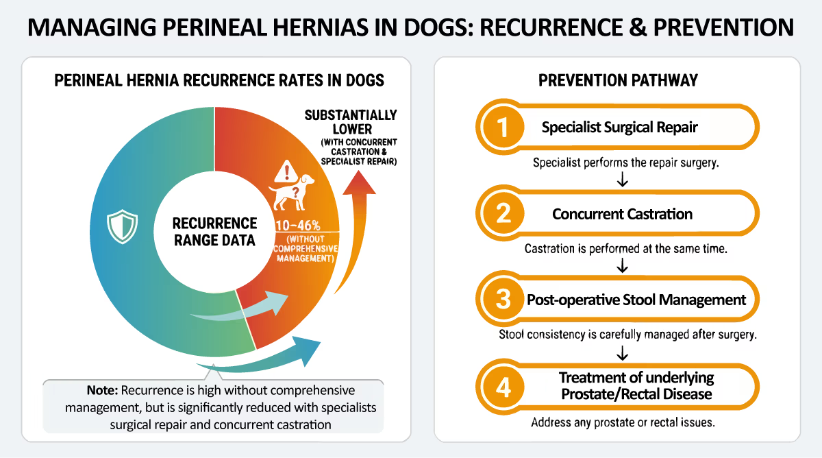
The prognosis for perineal hernia repair is generally good, but recurrence rates can range from 10% to 46% depending on the surgical technique used.
Postoperative complication rates are an important consideration when evaluating surgical outcomes, as different surgical methods and management strategies can influence the likelihood of complications following perineal hernia repair in dogs.
Overall Prognosis
Many dogs regain an excellent quality of life after surgical correction. Success depends on:
Surgical technique used
Surgeon experience
Whether castration was performed
Severity and chronicity of the hernia
Postoperative management of straining
Minor Postoperative Complications
Relatively common, usually self-limiting issues include:
Temporary swelling and bruising around the incision (swelling adjacent to the surgical site)
Mild bleeding or seroma formation
Discomfort when defecating for the first few days
Transient difficulty posturing
Minor wound redness
These typically resolve within 1–3 weeks with standard care.
Serious Complications
Less common but more concerning postoperative complications include:
Sciatic nerve injury during surgery can cause hindlimb lameness, knuckling, or weakness. Another postoperative nerve-related complication is nerve entrapment, which may cause pain, weakness, or neurological deficits soon after surgery. Careful surgical technique—avoiding deep suture placement near the sacrotuberous ligament—minimizes this risk.
| Complication | Description |
|---|---|
| Wound dehiscence | Breakdown of the surgical site closure |
| Surgical site infection | Bacterial contamination requiring treatment |
| Rectal prolapse | Protrusion of rectal tissue through the anus |
| Fistula formation | Abnormal tract between tissues |
| Nerve entrapment | Sciatic nerve damage causing hindlimb weakness or neurological deficits |
| Persistent constipation | Ongoing difficulty defecating |
Sciatic nerve injury during surgery can cause hindlimb lameness, knuckling, or weakness. Careful surgical technique—avoiding deep suture placement near the sacrotuberous ligament—minimizes this risk.
Fecal and Urinary Incontinence
Some dogs develop postoperative fecal incontinence, which may result from:
Pre-existing damage to the external anal sphincter
Intraoperative manipulation or nerve stretching
Chronic stretching of the anal sphincter before surgery
Most mild incontinence improves over weeks to months as inflammation resolves. However, some dogs—particularly those with bilateral disease or severe pre-existing sphincter damage—may have permanent changes.
Urinary incontinence is less common but can occur if bladder innervation is compromised.
Hernia Recurrence
Recurrence is the most significant long-term concern. Published recurrence rates vary widely:
Overall rates of 10–50% depending on technique, experience, and follow-up duration
Series using internal obturator muscle transposition report rates at the lower end (approximately 10–20%)
In one retrospective study examining clinical and surgical parameters in 84 dogs treated between 2008 and 2023, the recurrence rate was approximately 15%. Interestingly, dogs neutered before hernia development and those who developed postoperative fecal incontinence had higher odds of recurrence.
Factors that may increase recurrence risk:
Not performing castration at the time of repair
Bilateral disease with large defects
Very delayed surgical correction
Inadequate control of postoperative straining
Less robust surgical techniques
The clinical outcome varies, but most dogs with well-executed repairs and appropriate postoperative management enjoy years of comfortable, normal function.
Can Perineal Hernia Be Prevented, and How Do We Prevent Recurrence?
While there is no guaranteed way to prevent all perineal hernias, managing contributing factors can reduce risk throughout a dog’s life.
Primary Prevention Strategies
These measures may help reduce the likelihood of perineal hernia development:
Control constipation and diarrhea early: Address gastrointestinal issues promptly before chronic straining develops
Treat anal sac disease and colitis: Don’t ignore persistent scooting or straining
Manage urinary and prostatic problems: Early treatment of prostate disease in intact males reduces straining
Maintain healthy body condition: Regular exercise supports muscle health; avoid obesity
The Neutering Question
Historically, early castration was thought to prevent perineal hernia by reducing prostatic enlargement. The relationship is complex—while perineal hernias occur predominantly in intact males, suggesting a protective benefit of neutering, recent data on recurrence after repair show nuanced patterns.
The decision about when (or whether) to neuter should consider all health factors and be individualized with your veterinarian. For dogs diagnosed with perineal hernia, castration at the time of surgical repair remains strongly recommended.
Preventing Recurrence After Surgery
Secondary prevention—minimizing the chance of recurrence after repair—involves multiple strategies:
Surgical factors:
Using robust, modern techniques such as internal obturator transposition
Performing organopexies (colopexy, cystopexy) when indicated
Castrating intact males at the time of repair
Postoperative management:
Aggressive management of stool consistency during healing and long-term
Avoiding constipation through diet, hydration, and stool softeners
Treating any condition causing straining promptly
Practical Home-Care Measures
To minimize recurrence risk:
Continue high-fiber or prescription diet and stool softeners as recommended by your veterinarian
Ensure constant access to fresh water
Encourage regular toileting opportunities with multiple short walks daily
Monitor the perineal region for any new swelling or asymmetry
Watch for renewed straining, changes in tail carriage, or difficulty defecating
Seek veterinary review promptly if any concerns arise
For dogs with severe or recurrent disease, referral to a board-certified small-animal surgeon provides access to advanced techniques and specialized expertise that can reduce the risk of recurrence.
Living With a Dog After Perineal Hernia Repair
Most dogs return to comfortable, active lives after recovery. Some lifestyle adjustments and ongoing monitoring help ensure the best long-term outcome.
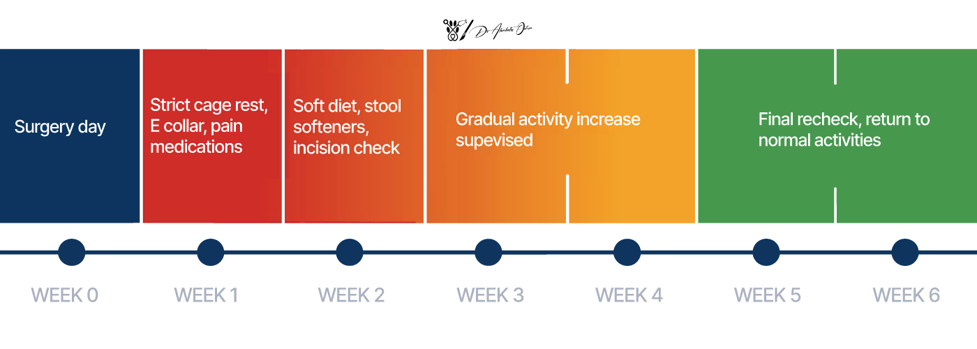
The First 2–3 Weeks
Strict home care during initial healing is essential:
Rest: Leash walks only; no running, jumping, or rough play
Protection: Elizabethan collar to prevent licking of the surgical site
Medication: Administer pain relief, antibiotics, and stool softeners exactly as prescribed
Monitoring: Check incisions daily for redness, swelling, discharge, or opening
Toileting Expectations
Encourage frequent short walks to stimulate gentle defecation and urination. Keep a simple log of whether your dog is passing stool and urine each day.
No defecation for 48 hours
Obvious straining or difficulty
Blood in stool or urine
Signs of pain or distress
Dietary Management
Soft, easily passed stools are ideal during recovery and often long-term:
Transition to high-fiber or veterinary gastrointestinal diets as directed
Avoid bones, rawhides, or very firm treats that can cause constipation
Many dogs benefit from adding water to kibble or feeding primarily wet food
Some dogs remain on mild stool softeners or fiber supplements indefinitely
Long-Term Considerations
Even after full recovery, remain vigilant:
Some dogs need ongoing dietary modification
Monitor for recurrence or development of a hernia on the opposite side
Age-related arthritis can make posturing to defecate more difficult—address this proactively
Regular veterinary check-ups allow early detection of any problems
Follow-Up Schedule
Plan for:
10–14 days: First recheck for suture removal and wound assessment
5–6 weeks: Additional review to confirm stable repair
Ongoing: As needed based on any concerns
Perineal Hernias and Quality of Life
Perineal hernias can have a profound impact on the quality of life for affected dogs, particularly older intact male dogs, who are at the highest risk. When the pelvic diaphragm weakens or fails, abdominal organs may herniate into the perineal region, leading to a cascade of clinical signs that disrupt a dog’s daily comfort and well-being.
Dogs with perineal hernia often experience persistent perineal swelling, which can make sitting, walking, and especially defecating uncomfortable. Chronic constipation and straining are common because herniated tissue can distort the rectal wall and impede normal stool passage. This not only causes discomfort but can also lead to more serious issues, such as rectal prolapse or fecal incontinence, further diminishing a dog’s quality of life.
Urinary problems are another significant concern. If the urinary bladder becomes trapped within the hernia, dogs may develop urinary obstruction or even urinary incontinence. These complications can be distressing for both the dog and their owner, and in severe cases, may become life-threatening emergencies requiring immediate intervention.
Surgical treatment is the cornerstone of restoring quality of life in dogs with perineal hernia. Perineal hernia repair, particularly with advanced techniques such as internal obturator muscle transposition, aims to reconstruct the pelvic diaphragm and prevent further herniation of abdominal organs. In some cases, surgical mesh may be used to reinforce the repair, especially when the defect is large or the tissue quality is poor. These procedures are designed to relieve clinical signs, restore normal function, and reduce the risk of recurrence.
Postoperative care plays a critical role in ensuring a successful outcome. Pain management, careful monitoring for postoperative complications such as surgical site infection or wound dehiscence, and strategies to prevent constipation are all essential. While most dogs recover well, some may develop postoperative fecal incontinence or other complications that require ongoing management. Regular follow-up with your veterinary surgeon is important for monitoring healing, addressing any issues promptly, and supporting your dog’s long-term well-being.
With timely surgical intervention and diligent postoperative care, most dogs experience a dramatic improvement in their ability to defecate, reduced discomfort, and a return to a more active, comfortable life. Although hernia recurrence and postoperative complications are possible, especially in cases with severe tissue weakness or delayed treatment, most dogs experience a significantly improved quality of life following perineal hernia repair.
FAQ: Perineal Hernia in Dogs
Is my dog in pain with a perineal hernia?
Many dogs experience mild discomfort or only notice difficulty when straining to defecate, especially in the early stages. However, pain can become severe if the urinary bladder or intestine becomes trapped within the hernia. Dogs with acute bladder retroflexion or strangulated bowel are often distressed, vocalizing, and may collapse. Early elective therapies and surgical treatment, before complications develop, significantly improve your dog’s comfort.
How much does perineal hernia surgery cost, and will insurance help?
Costs vary considerably depending on geographic region, whether treatment occurs at a primary care practice or a referral center, and case complexity (emergency vs. elective, unilateral vs. bilateral, presence of bladder or intestinal involvement). Referral centers with board-certified surgeons typically charge more but offer advanced expertise. Many pet insurance policies cover perineal hernia surgery if the condition is not pre-existing. Contact your insurer before treatment to understand your specific coverage.
Will my dog need surgery on the other side as well?
Many dogs eventually develop a hernia on the contralateral side, particularly if they remain intact after initial diagnosis. Studies suggest that bilateral involvement is present in at least 30% of cases at first presentation, and more dogs develop it over time. Careful surgical technique, castration at the time of repair, and lifelong management of straining can reduce—but not eliminate—this risk.
Is there an age at which my dog is too old for repair?
Age alone is not an absolute limit for surgery. Overall health, heart and kidney function, and quality-of-life goals are more important considerations. Many senior dogs do very well with appropriate pre-anesthetic assessment, tailored anesthetic protocols, and careful perioperative care. Discuss your individual dog’s situation with your veterinarian or surgeon to make an informed decision.
What happens if I decide against surgery?
Without surgical correction, perineal hernias typically enlarge over time. Defecation becomes progressively more difficult, and the risk of life-threatening complications—such as bladder herniation with urinary obstruction or intestinal strangulation—remains constant. Palliative medical management can provide temporary relief and buy time, but it cannot repair the structural weakness. Most vet surg specialists recommend surgery as the only realistic option for long-term quality of life.