Quick Facts for Pet Owners
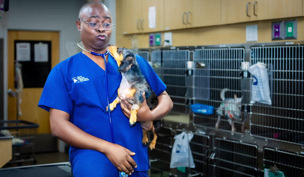
Chylothorax is a rare but serious condition in dogs and cats where lymphatic fluid rich in fat accumulates around the lungs inside the chest cavity. This fluid buildup compresses the lungs, causing difficulty breathing that can become life-threatening without prompt veterinary care. If your pet is breathing rapidly, struggling to catch their breath, or refusing to lie down, immediate veterinary attention is essential.
This guide is intended for pet owners and veterinary professionals seeking to understand the causes, diagnosis, treatment, and prognosis of chylothorax in dogs and cats. Early recognition and intervention are crucial for improving outcomes in affected pets.
-
Chylothorax is uncommon but potentially life-threatening if left untreated
-
Most cases in dogs and cats are idiopathic (no identifiable cause) or secondary to heart disease, lymphatic disorders, or cancer
-
Urgent veterinary evaluation is critical if your pet shows labored breathing, open-mouth breathing, or chest pain
-
Treatment options range from drainage and dietary changes to medication and surgical intervention
-
This information is specific to dogs and cats—human chylothorax has different causes and management approaches
-
Early involvement of a veterinary surgeon or specialist improves outcomes significantly
What Is Chylothorax in Dogs and Cats?
Chylothorax occurs when chyle—a milky-white lymphatic fluid rich in triglycerides and lymphocytes—accumulates within the pleural space surrounding the lungs. This happens when the thoracic duct or its branches leak lymph fluid into the chest cavity instead of returning it to the bloodstream. The result is a form of pleural effusion that progressively restricts lung expansion and impairs breathing. Chyle specifically collects in the pleural cavity, leading to respiratory distress and requiring prompt diagnosis and management.
Chyle itself originates from the intestines. When your dog or cat digests dietary fat, the small intestine absorbs long-chain fatty acids into specialized lymphatic vessels called lacteals. Chyle is formed in the small intestine during the digestion of fatty foods and contains triglycerides, lymphocytes, proteins, and fat-soluble vitamins. These converge into the cisterna chyli near the abdomen and then flow through the thoracic duct, ultimately draining into the venous system near the heart. When this pathway is disrupted—whether by trauma, obstruction, or unknown causes—chylous fluid escapes into the surrounding pleural space and accumulates. In rare cases, chyle or lymphatic fluid from the peritoneal cavity can migrate into the thoracic (pleural) cavity through transdiaphragmatic defects or increased intra-abdominal pressure.
Chylothorax differs from other types of pleural effusion:
-
Hemothorax contains blood from trauma or bleeding disorders
-
Pyothorax consists of pus from bacterial infection
-
Transudates and exudates may result from heart failure, liver disease, or cancer
-
Chylothorax specifically involves triglyceride-rich lymphatic fluid from the thoracic duct
This condition is seen in both dogs and cats worldwide. Certain breeds appear overrepresented in veterinary literature, including Afghan Hounds, Shiba Inus, and Siamese-type cats, though any breed can be affected.
Relevant Thoracic and Lymphatic Anatomy
Understanding the lymphatic system anatomy is essential for both diagnosing and treating chylothorax in dogs and cats. The cisterna chyli originates near the second and third lumbar vertebrae (L2–L3), collecting lymphatic drainage from the intestines and abdominal organs. From there, the thoracic duct ascends through the aortic hiatus of the diaphragm and travels along the dorsal mediastinum, typically on the right side of the aorta initially.
At approximately the level of the fifth to seventh thoracic vertebrae (T5–T7), the duct crosses from right to left before terminating at the junction of the jugular veins and subclavian veins near the cranial vena cava. This crossover point explains why the side of chylous effusion can vary. Right-sided chylothorax tends to occur when disruption happens caudal to the crossover, while left-sided or bilateral chylothorax results from cranial disruption.
Key anatomical points for clinicians:
-
The thoracic duct wall is thin and fragile, making it susceptible to trauma and surgical injury
-
Multiple collateral lymphatic branches and accessory ducts exist, creating significant anatomical variation between individual animals
-
These variations directly affect surgical planning for thoracic duct ligation
-
Preoperative lymphatic imaging can help identify duct anatomy before surgery
-
The duct’s small diameter and variable course make complete ligation challenging
-
Advanced imaging modalities such as CT, ultrasound, or MRI can also assess the chest wall for abnormalities, such as injuries, tumors, or malformations, that may contribute to or complicate chylothorax by affecting the pleural cavity or lymphatic drainage
Documented anatomical variations in canine and feline imaging studies show that some animals have multiple parallel ducts or unusual termination points. This variability is why surgeons often perform mass ligation of the tissue surrounding the expected duct location rather than attempting to isolate a single vessel.
Causes and Risk Factors (Etiology)
Chylothorax in dogs and cats can result from a variety of causes, including traumatic injury, cancer, congenital defects, lymphatic disorders, and heart issues. It is associated with a range of thoracic diseases, both malignant and benign.
In dogs and cats, chylothorax is broadly classified into three categories: idiopathic chylothorax (no identifiable cause), nontraumatic chylothorax secondary to underlying disease, and traumatic chylothorax from injury or iatrogenic causes. Nontraumatic chylothorax can result from congenital, neoplastic, infectious, or idiopathic causes. In contrast, traumatic chylothorax results from iatrogenic injury during surgery or from non-surgical trauma such as blunt chest injuries or penetrating wounds. A thorough workup to identify the underlying cause is critical because prognosis depends heavily on etiology.
Common Causes in Dogs
-
Idiopathic chylothorax (most common, particularly in Afghan Hounds and Shiba Inus)
-
Cranial mediastinal masses, especially lymphoma and thymoma
-
Pericardial effusion and constrictive pericarditis
-
Right-sided heart failure causing thoracic duct obstruction through elevated venous pressure
-
Thoracic duct thrombus
-
Diaphragmatic hernia
-
Fungal granulomas affecting mediastinal lymph nodes
-
Post-surgical leaks following thoracotomy or PDA ligation
Common Causes in Cats
-
Idiopathic chylothorax
-
Cardiomyopathies, notably hypertrophic cardiomyopathy and restrictive cardiomyopathy
-
Mediastinal lymphoma (malignant chylothorax)
-
Heartworm disease in endemic regions
-
Thoracic trauma
-
Post-pericardiectomy or other thoracic procedures
Idiopathic chylothorax remains frustratingly common in both species. Despite extensive imaging and testing, no underlying disease can be identified in many patients. Some researchers speculate that subtle lymphangiectasia, congenital abnormalities of lymphatic vessels, or microthrombi not visible on routine imaging may be responsible.
Traumatic chylothorax and iatrogenic injury can follow blunt trauma (such as being hit by a car or high-rise falls in cats), penetrating wounds, or thoracic surgery. In human medicine, postoperative chylothorax frequently follows cardiothoracic surgery procedures like coronary artery bypass surgery, coronary artery bypass grafting, lung cancer surgery, lung resection, or thoracic aneurysm repair. While some of these specific surgeries are rarely performed in veterinary patients, the principle remains the same: any procedure near the thoracic duct can cause lymph leakage. Postoperative complications typically appear within days to a few weeks after the procedure.
Congenital chylothorax associated with lymphatic malformations is recognized in young animals and parallels patterns of congenital heart disease in neonates. These cases may present very early in life and can be associated with other lymphatic disorders such as chylous ascites.
Pathophysiology
Chylothorax occurs when there is disruption, obstruction, or increased pressure within the thoracic duct or lymphatic system of dogs and cats, causing lymphatic fluid to leak into the chest cavity rather than return to the venous circulation.
The main mechanisms include:
Direct duct rupture from trauma, surgical injury, or penetrating wounds
Functional obstruction from mass effect (tumors pressing on the duct) or thrombus formation
Elevated venous and lymphatic pressures from right-sided heart failure, pericardial disease, or venous fat hemorrhage patterns
Congenital or degenerative malformations of thoracic lymphatics leading to lymphangiectasia and spontaneous leakage
The composition of chyle explains many systemic consequences:
High triglyceride content (from absorbed long-chain fatty acids)
High lymphocyte count (primarily T cells recirculating through gut-associated lymphoid tissue)
Fat-soluble vitamins (A, D, E, K)
Albumin and immunoglobulins
Essential fatty acids
Chronic loss of these components leads to progressive malnutrition, hypoproteinemia, and immunosuppression. Dogs and cats with persistent drainage of chyle become increasingly vulnerable to infections due to lymphopenia and loss of white blood cells.
The mechanical effects of persistent effusion are equally damaging:
Atelectasis (lung collapse) from external compression
Reduced lung compliance
Hypoxemia and tachypnea
Progressive exercise intolerance
In chronic cases, restrictive pleuritis develops as fibrin and collagen deposit on the pleural surface
This chronic pleural fibrosis (“fibrosing pleuritis”) represents one of the most serious complications. Thick fibrous tissue encases the lungs, preventing normal expansion even after fluid is removed. Once established, this fibrosis is irreversible and dramatically worsens surgical outcomes.
Clinical Signs and Physical Examination Findings
The severity of clinical signs in dogs and cats correlates primarily with the volume and rate of chyle accumulation rather than the specific underlying cause. A slowly developing effusion may initially present with subtle signs, whereas rapid accumulation can lead to acute respiratory distress.
Common Clinical Signs
Tachypnea (increased respiratory rate)
Open-mouth breathing (especially concerning in cats)
Increased abdominal effort during breathing
Exercise intolerance
Lethargy and reduced activity
Decreased appetite
Progressive weight loss
Muscle wasting in chronic cases
Poor coat quality and dull fur
Coughing is variably present in dogs with chylothorax but uncommon in cats. Many cats instead present with quiet but rapid breathing and a reluctance to lie on one side. Some cats may hide or become unusually inactive before showing obvious respiratory distress.
Physical Examination Findings
Restrictive breathing pattern with short, shallow breaths
Muffled heart and lung sounds ventrally (where fluid accumulates)
Normal or increased breath sounds dorsally
Cyanotic (blue-tinged) mucous membranes in severe cases
Weak peripheral pulses in advanced cases
Possible palpable cranial mediastinal mass on thoracic palpation or imaging
Clinical vignette—dog: A 6-year-old Afghan Hound presents with three weeks of progressive exercise intolerance and increased respiratory effort. The owner notes the dog now refuses to lie flat and prefers sternal recumbency. Auscultation reveals muffled lung sounds bilaterally in the ventral thorax.
Clinical vignette—cat: A 10-year-old Siamese cat is brought in for acute open-mouth breathing that started overnight. The cat has been hiding more than usual for the past week. Physical examination reveals tachypnea, abdominal breathing effort, and diminished breath sounds on the right side.
Some animals present in acute respiratory crisis require immediate oxygen supplementation and emergency thoracocentesis before any diagnostic workup can proceed.
Diagnostic Approach
The diagnosis of chylothorax in dogs and cats requires both confirming the presence of chyle in the pleural space and systematically searching for an underlying cause. The diagnostic sequence typically proceeds through stabilization, imaging, thoracocentesis with pleural fluid analysis, and advanced testing as indicated.
Emergency Stabilization
Before pursuing extensive diagnostics in a dyspneic animal, stabilization takes priority:
Oxygen supplementation via flow-by, mask, or oxygen cage
Gentle handling with minimal stress
Thoracocentesis to remove enough fluid to ease breathing if respiratory distress is severe
Avoid forcing the animal into lateral recumbency, which may worsen respiratory compromise
Imaging
After partial stabilization, imaging helps characterize the effusion and identify potential causes:
Thoracic radiographs (chest radiographs or chest x ray) after partial fluid removal reveal pleural fissure lines, lung lobe retraction, and soft-tissue opacity. Pre-drainage films often underestimate underlying disease because fluid obscures the lungs and mediastinum.
Thoracic ultrasound provides rapid assessment of pleural fluid volume, guides safe thoracocentesis, and can identify cranial mediastinal masses, pericardial effusion, and cardiac abnormalities.
Computed tomography (CT) with contrast enhancement offers superior detection of mediastinal masses, lung lobe torsion, hernias, and thoracic duct anatomy.
Lymphatic imaging techniques (conventional lymphangiography, CT lymphangiography, or MR lymphangiography) can map the site of lymphatic leakage when available at referral centers.
Pleural Fluid Analysis
Thoracocentesis and comprehensive pleural fluid analysis are the cornerstones for confirming chylothorax:
Gross appearance: Often milky-white, but may be serosanguineous or pink-tinged in fasting animals or those with concurrent hemorrhage
Cytology: Small lymphocyte predominance initially; may become neutrophil-predominant with chronic inflammation
Biochemistry: A pleural fluid triglyceride concentration greater than the simultaneous serum triglyceride concentration is diagnostic. In humans, pleural fluid triglyceride >110 mg dl strongly supports chylothorax; veterinary diagnosis relies on the same principle.
Cholesterol: Typically lower in chylous fluid than in pseudochylous effusions
The triglyceride comparison is essential because not all chylous fluids appear milky. Fasting animals or those with concurrent hemorrhage may have a misleading fluid appearance.
Additional Diagnostics
To diagnose chylothorax fully and identify causes, additional testing includes:
Echocardiography to evaluate for cardiomyopathy, valvular disease, heart failure, and pericardial disease
Fine-needle aspirates of mediastinal masses or enlarged lymph nodes if identified
Routine bloodwork (CBC, biochemistry, total proteins) to assess nutritional and immunologic impact and prepare for anesthesia
Heartworm testing in endemic regions for cats
Differential Diagnosis in Dogs and Cats
Many diseases cause pleural effusion with similar respiratory signs, and differentiating them is essential for appropriate therapy. A chylothorax diagnosed through proper fluid analysis must still be distinguished from other effusion types.
Other Pleural Effusions in Small Animals
| Effusion Type | Appearance | Predominant Cells | Triglycerides | Cholesterol | Common Causes |
|---|---|---|---|---|---|
| Chylothorax | Milky-white to pink | Lymphocytes (early) | Higher than serum | Lower than serum | Idiopathic, heart disease, neoplasia, trauma |
| Pyothorax | Turbid, foul-smelling | Degenerate neutrophils, bacteria | Normal | Variable | Bacterial infection, foreign body, bite wounds |
| Hemothorax | Red, bloody | Red blood cells | Normal | Normal | Trauma, coagulopathy, neoplasia |
| Transudates | Clear to straw-colored | Few cells | Low | Low | Heart failure, hypoproteinemia |
| Neoplastic effusion | Variable | Atypical/neoplastic cells | Variable | Variable | Lymphoma, carcinoma, mesothelioma |
| Pseudochylothorax | Milky (cholesterol crystals) | Variable | Lower than serum | Higher than serum | Chronic inflammatory effusions |
Key differentiating features:
Pyothorax typically has a foul odor, neutrophilic exudate with bacteria visible on cytology, and affected animals are often febrile
Neoplastic effusions may show atypical cells on cytology, though not always
Pseudochylothorax (rare in small animals) shows high cholesterol with lower triglycerides and cholesterol crystals on cytology
Multiple concurrent processes may coexist. An animal with chylothorax secondary to mediastinal lymphoma may have both chylous and neoplastic components in the effusion. Fluid analysis must always be interpreted in conjunction with imaging and clinical history.
Treatment and Management Options
The management of chylothorax in dogs and cats follows a stepwise approach: stabilize the patient, drain accumulated fluid, identify and treat the underlying cause when possible, and implement dietary and medical therapies. To effectively treat chylothorax, a multidisciplinary approach is often required, incorporating both nonsurgical and surgical interventions.
Treatment for chylothorax includes conservative methods like dietary changes and chest drains, progressing to interventional procedures if necessary. In dietary management, changes may involve a low-fat diet or medium-chain triglyceride (MCT) diet to reduce chyle production, as MCTs are absorbed directly into the portal circulation and do not enter the lymphatic system.
Medical therapies may include somatostatin and octreotide, which reduce chyle formation and promote closure of thoracic duct leaks. In severe cases, Total Parenteral Nutrition (TPN) may be used to provide nutrition intravenously and stop digestive chyle production.
Fluid drainage strategies often begin with intermittent therapeutic thoracentesis, which is commonly used in the initial management of chylothorax to alleviate dyspnea caused by pleural fluid.
For patients who persistently reaccumulate fluid despite dietary modifications and repeated thoracenteses, pleurodesis is indicated. Surgical intervention is considered when conservative measures fail or when a surgically correctable cause is identified.
Emergency Care
Oxygen therapy via flow-by, mask, or oxygen cage
Therapeutic thoracocentesis to remove sufficient fluid to ease breathing
Hospitalization and monitoring for severely compromised patients
Intravenous fluid support if needed, with caution to avoid fluid overload
Fluid Drainage Strategies
Intermittent thoracocentesis may be adequate for slow-accumulating effusions
Temporary chest tube (thoracostomy tube) placement allows repeated drainage without multiple needle punctures
Balance must be struck between symptom relief and risks of repeated anesthesia, infection, and ongoing protein/lymphocyte loss
Dietary Management
Ultra-low-fat diet or a commercial veterinary low-fat formula reduces chyle production
Medium-chain triglyceride (MCT) supplements are sometimes used, though evidence in dogs and cats is less robust than in humans
Cats have strict nutritional requirements; prolonged fat restriction requires close oversight to prevent hepatic lipidosis and ensure adequate taurine intake
Medical Therapies
Rutin (a benzopyrone flavonoid) is commonly used in veterinary practice, typically dosed several times daily. The proposed mechanism involves enhancing macromolecule clearance and stimulating macrophage activity, though robust evidence is limited.
Octreotide or somatostatin analogs have been used in select refractory cases, extrapolated from human experience with reducing lymphatic flow
Diuretics alone do not resolve chylothorax and may worsen dehydration and electrolyte imbalance; they serve only as adjuncts when cardiac disease is present
Surgical Indications
Consider surgical treatment when:
A surgically correctable underlying cause is identified
Idiopathic chylothorax fails to respond to 4–8 weeks of medical and dietary management
Daily drainage volumes remain high
Progressive weight loss or declining body condition occurs despite conservative therapy
Prolonged conservative management risks allowing irreversible pleural fibrosis to develop, dramatically worsening long-term outcomes. The timing of surgery should be discussed with owners early.
Surgical Management in Dogs and Cats
Surgery offers the best chance of long-term control for many dogs and cats with idiopathic or secondary chylothorax, particularly when performed before advanced pleural fibrosis develops.
Thoracic Duct Ligation (TDL)
Thoracic duct ligation remains the cornerstone surgical procedure. The approach typically involves:
Right-sided intercostal thoracotomy at the 8th–10th intercostal space, or thoracoscopic approach
Intraoperative lymphangiography using cream or lipid infusion to visualize the duct and lymphatic vessels
Mass ligation of tissue in the duct region to encompass anatomical variations
Application of multiple ligatures or vascular clips
TDL alone in dogs historically achieves approximately 50–60% success. Success rates improve significantly when combined with adjunctive procedures.
Combined Procedures
Subtotal pericardectomy reduces right atrial pressure and facilitates alternative lymphatic drainage pathways
Cisterna chyli ablation redirects lymphatic flow away from the thoracic duct
In cats, combined TDL with subtotal pericardectomy has shown good outcomes in published case series, though data are more limited than for dogs
Managing Advanced Cases
For animals with established restrictive pleuritis:
Decortication (pleural stripping) may be needed to remove fibrous tissue encasing the lungs
These procedures carry higher morbidity and have variable outcomes
Prognosis is significantly more guarded in chronic, fibrotic cases
Perioperative Care
Chest drain placement for postoperative monitoring and drainage
Multimodal analgesia
Nutritional support, especially in debilitated patients
Monitoring of effusion volume and character
Re-evaluation of diet following surgery
Surgical risks include hemorrhage, failure to identify all lymphatic branches, persistent effusion requiring reoperation, and anesthetic complications in already compromised patients. Experience matters significantly—outcomes are generally better at centers with thoracic surgeons experienced in these procedures.
Interventional Radiology and Emerging Techniques
Although still limited in availability relative to human medical centers, minimally invasive lymphatic interventions are emerging options at veterinary referral hospitals for dogs and, occasionally, cats with refractory chylothorax.
Thoracic Duct Embolization (TDE)
Thoracic duct embolization involves:
Catheterization of the thoracic duct via the mesenteric lymphatic or cisterna chyli approach
Occlusion using coils, glue, or other embolic agents to stop chyle leakage
Performed under fluoroscopic guidance with lymphatic imaging
Veterinary case reports describe successful thoracic duct embolization in dogs; however, the technique requires specialized interventional radiology equipment and expertise that are not available at most practices.
Indications for Interventional Approaches
Patients who have failed conventional surgical treatment
Poor surgical candidates due to anesthetic risk or concurrent disease
Cases where advanced imaging can precisely localize the leak site
Potential Complications
Fat embolism
Local tissue swelling
Contrast reactions
Technical failure requiring conversion to surgery
Comparison with Surgery
| Factor | Surgical TDL | Thoracic Duct Embolization |
|---|---|---|
| Invasiveness | Moderate to high | Lower |
| Availability | Referral surgical centers | Limited specialized centers |
| Recovery time | Days to weeks | Generally shorter |
| Technical requirements | Surgical expertise | Interventional radiology suite |
As the veterinary evidence base grows, percutaneous lymphatic interventions may increasingly complement traditional surgical approaches.
Nutritional and Medical Management in Detail
Regardless of whether surgery is performed, nutritional support and medical therapy are vital to maintain body condition and immune function in dogs and cats with chronic chylothorax.
Dietary Principles in Dogs
Commercial low-fat veterinary diets (ideally <10% fat on a dry matter basis)
Careful treat selection—avoid high-fat table scraps and fatty treats
Monitor body weight and muscle condition regularly
Ensure the diet remains complete and balanced despite fat restriction
Consider MCT supplementation cautiously, as evidence supporting its use is limited
Special Considerations in Cats
Higher risk of hepatic lipidosis with inadequate caloric intake
Strict taurine requirements must be met
Lesser tolerance for prolonged fat restriction than dogs
Close oversight by a veterinary nutritionist is recommended
More frequent monitoring of appetite and food intake
Rutin Therapy
Rutin, a plant-derived benzopyrone, is commonly prescribed for canine and feline chylothorax:
Proposed mechanism: stimulates macrophage uptake of protein and promotes lymphatic drainage
Typical dosing: administered two to three times daily (dose varies by patient size)
Time to effect: weeks of treatment before improvement may be seen
Evidence: mixed results in small studies; widely used empirically but not definitively proven
Other Medical Options
Octreotide infusions have been reported in refractory cases
Cost and limited veterinary data restrict routine use
These agents are considered when conventional treatments fail
Monitoring Parameters
Regular reassessment should include:
Serial body weight measurements
Body condition score and muscle condition score evaluation
Serum albumin and globulin levels
Fat-soluble vitamin status in long-standing effusions
Complete blood count to monitor lymphocyte levels
Home Care, Activity, and What Owners Should Avoid
Most dogs and cats with chylothorax require ongoing monitoring at home even after discharge from the hospital or following surgery. Understanding proper home management helps prevent complications and identifies problems early.
Activity Restrictions
Avoid intense exercise while effusion is present or during early recovery
Prevent overheating and stressful situations that increase respiratory effort
Short, calm leash walks are generally acceptable
Cage rest may be recommended immediately after surgery
Dietary Compliance
Feed only the veterinary-prescribed low-fat diet
Strictly avoid table scraps and high-fat treats
Unlike human chylothorax management, complete fasting and intravenous nutrition are rarely used long-term at home in small animals
Ensure fresh water is always available
Warning Signs Requiring Urgent Veterinary Care
Increased respiratory rate at rest (consistently over 30–35 breaths per minute)
Open-mouth breathing
Obvious effort to breathe (abdominal heaving)
Cyanotic (blue or purple) tongue or gums
Collapse or sudden extreme lethargy
Complete refusal to eat for more than 24 hours
Caring for Chest Drains at Home
If your pet is discharged with a temporary thoracostomy tube:
Keep bandages clean and dry
Prevent chewing or pulling at the tube (Elizabethan collar may be needed)
Check regularly for air leaks, excessive drainage, or disconnection
Note the volume and color of any drained fluid
Contact your veterinary clinic immediately if anything seems abnormal
Prognosis and Long-Term Outlook
The prognosis for chylothorax in dogs and cats varies considerably and depends on the underlying cause, the duration before diagnosis, and the response to treatment. Understanding what to expect helps owners make informed decisions about their pet’s care.
Favorable Prognosis
Traumatic or iatrogenic chylothorax often resolves well if the underlying injury can be controlled and the chyle leak stops within days to weeks
Some post-surgical leaks resolve spontaneously with conservative management
Guarded but Acceptable Prognosis
Idiopathic chylothorax carries more uncertainty, but published surgical series report long-term control in the majority of dogs and many cats following thoracic duct ligation combined with adjunct procedures
Success depends significantly on timing—earlier intervention before pleural fibrosis develops improves outcomes
Poor Prognosis
Chylothorax secondary to malignant neoplasia (such as mediastinal lymphoma) or advanced cardiac disease has a poorer outlook because the underlying condition may not be curable
The key factor influencing prognosis in these cases is the treatability of the primary disease
Consequences of Delayed or No Treatment
Chronic, untreated chylothorax leads to irreversible pleural fibrosis
Permanent restrictive lung disease dramatically worsens quality of life
Progressive malnutrition, immunosuppression, and debilitation occur
Quality-of-Life Considerations
Owners should consider:
Need for repeated thoracocentesis if surgery is not pursued
Financial impact of ongoing treatment versus surgical intervention
Time commitment for frequent veterinary visits and home monitoring
In refractory cases that do not respond to treatment, humane euthanasia may be the most compassionate choice
Published veterinary studies suggest that median survival times vary widely by etiology and treatment approach. Dogs with idiopathic chylothorax treated surgically often survive years with a good quality of life, while those with underlying malignancy may survive only weeks to months.
Complications and Comorbidities
The disease process and its treatment can lead to multiple systemic complications in dogs and cats. Recognizing and managing these complications improves outcomes.
Nutritional Complications
Loss of fats, proteins, and fat-soluble vitamins through chyle
Progressive weight loss and muscle wasting
Poor wound healing after surgery
Dull, dry coat quality
Immunologic Effects
Chronic loss of lymphocytes and immunoglobulins in chyle
Increased susceptibility to respiratory infections
Higher risk of wound infections following surgery
Potential for sepsis in debilitated patients
Respiratory Complications
Chronic atelectasis from persistent effusion
Restrictive pleuritis, limiting lung expansion
Pulmonary hypertension in severe cases
Secondary lung infections from decreased lung expansion and ventilation
Treatment-Related Complications
Risks associated with repeated anesthesia in compromised patients
Iatrogenic pneumothorax from thoracocentesis
Infection or blockage of chest tube drains
Bleeding during surgery
Chyle leak persistence after thoracic duct ligation
Infectious chylothorax can occur secondary to contamination during procedures
Minimizing Complications
Meticulous aseptic technique during all procedures
Judicious timing of interventions based on patient stability
Early nutritional support before marked debilitation occurs
Regular re-evaluation to detect problems promptly
Subspecialty consultation for complex cases
When Should Owners Seek Veterinary Help or Specialist Referral?
Any dog or cat with labored breathing, rapid breathing at rest, or open-mouth breathing should be seen by a veterinarian immediately, regardless of whether a diagnosis has already been established.
Emergency Warning Signs
Pronounced abdominal movement when breathing
Inability to lie down comfortably
Restlessness and constant position changes due to air hunger
Blue or purple discoloration of the tongue or gums
Collapse or inability to stand
Extreme weakness or unresponsiveness
When to Recheck Animals Already Diagnosed
Resting respiratory rate increases above baseline
Cough develops or worsens
Lethargy increases beyond normal rest
Appetite declines for more than 24 hours
Any sudden change in breathing pattern
Indications for Specialist Referral
General practitioners should consider referral to a board-certified surgeon or internal medicine specialist when:
Chylous effusion persists beyond a few weeks despite conservative therapy
Drainage volumes remain high
Effusion recurs after initial resolution following trauma
Cranial mediastinal mass is suspected and requires advanced imaging and possible surgical biopsy
Thoracic duct ligation or other surgical intervention is being considered
Earlier referral in idiopathic cases improves surgical planning, allows access to lymphatic imaging when available, and generally leads to better long-term outcomes. Waiting until pleural fibrosis has developed significantly limits treatment options.
Questions Owners May Want to Ask Their Veterinarian
Informed owners can more effectively partner in decision-making regarding dogs and cats with chylothorax. Consider asking these questions during appointments:
About Diagnosis
“What tests have confirmed that this is chylothorax rather than another type of pleural effusion?”
“Have we ruled out heart disease, cancer, and diaphragmatic hernia as possible causes?”
“Would additional imaging like CT or echocardiography help identify an underlying cause?”
About Treatment
“What are the realistic goals of medical management alone in my dog/cat?”
“At what point would you recommend surgery or referral to a surgical specialist?”
“What are the potential benefits and risks of thoracic duct ligation in my pet’s case?”
“How many similar cases have you or the referral surgeon treated?”
About Prognosis and Home Care
“What should I feed at home and for how long?”
“What signs at home should trigger an emergency visit?”
“What is the expected prognosis and quality of life with and without surgery?”
“How often will my pet need to return for fluid drainage?”
Take notes during consultations and request clarification on any points that are uncertain. Understanding the diagnostic and therapeutic strategies helps you advocate effectively for your pet.
Coordination of Care and Veterinary Team Roles
Optimal management of chylothorax in dogs and cats requires an interprofessional veterinary team. This collaborative approach mirrors human medical models but is adapted to small-animal practice.
Primary Care Veterinarian
Early recognition of respiratory distress
Initial stabilization and fluid drainage
Preliminary diagnostics, including radiographs and fluid analysis
Timely referral to specialists when indicated
Long-term monitoring and medication management after specialist treatment
Board-Certified Surgeons
Thoracic duct ligation and combined procedures
Pericardectomy and cisterna chyli ablation
Management of restrictive pleuritis and decortication when needed
Expertise in treating chylothorax in complex cases
Coordination with anesthesiologists for safe procedures
Internal Medicine Specialists
Advanced imaging interpretation
Comprehensive cardiac evaluation
Management of underlying diseases such as cardiomyopathy or lymphoma
Long-term medical management coordination
Additional Team Members
Veterinary anesthesiologists ensure safe anesthesia in patients with compromised respiratory function
Critical care specialists manage severely ill patients requiring intensive monitoring
Radiologists provide expert CT and MR interpretation and may perform lymphatic imaging
Veterinary nutritionists design customized low-fat diets, particularly important for cats
Communication and Expectations
Clear communication with owners about goals of care, costs, potential need for repeated procedures, and long-term monitoring is essential. Outcomes improve significantly when cases are managed in a coordinated manner at centers experienced in small-animal chylothorax.
Key Takeaways
Chylothorax is a rare but serious condition requiring prompt diagnosis and treatment
The diagnosis of chylothorax depends on confirming elevated pleural fluid triglycerides compared to serum
A full workup to identify the underlying cause determines appropriate treatment and prognosis
Conservative management with diet and medication may help, but often fails in idiopathic cases
Surgical intervention with thoracic duct ligation and combined procedures offers the best chance for long-term resolution
Thoracic duct embolization is an emerging minimally invasive option at specialized centers
Early specialist referral—before pleural fibrosis develops—improves outcomes
Yellow nail syndrome and subcutaneous lesions are not typical features in veterinary chylothorax, but are reported in some human lymphatic syndromes
Chylothorax and associated outcomes vary considerably between patients, but with appropriate treatment, many dogs and cats can return to comfortable, active lives. If your pet is showing signs of breathing difficulty, don’t wait—contact your veterinarian immediately. Early recognition and specialist involvement remain the most reliable path to a positive outcome.